The most important reasons for performing echocardiography
Physicians use esophageal echocardiography for sensitive surgeries and important treatment decisions, especially when other methods do not provide enough information. Echo Ego is a key tool in the management of heart disease and helps to maintain the patient's health in the best possible way.
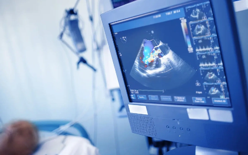
- Providing very high resolution images due to the proximity of the transducer to the heart
- Cardiac evaluation before major surgeries or immediately after open heart surgery
- ensuring proper heart and valve function
- exact examination of heart valve problems such as aortic and mitral valve stenosis or insufficiency
- Important role in identifying intracardiac blood clots (especially in patients with atrial fibrillation or after stroke)
- providing sufficient and more detailed information about the structure or function of the heart than other imaging methods
- Diagnosis of structural abnormalities of the heart such as inter-atrial or ventricular holes
- The possibility of accurate diagnosis of complex diseases and appropriate treatment
- effective in checking the performance of artificial valves
- Effective role in identifying abscesses or infectious masses in patients suspected of endocarditis (heart valve infection)
In what cases is echocardiography performed?
Echo of the heart or transesophageal echocardiography (TEE) is used when echocardiography from the chest cannot provide clear and sufficient images.
Know the types of esophageal echocardiography...
Each type of echocardiography has its own use and advantages, and the cardiologist will choose the right type according to your needs. Familiarity with these types can help to better understand the applications of this advanced method.
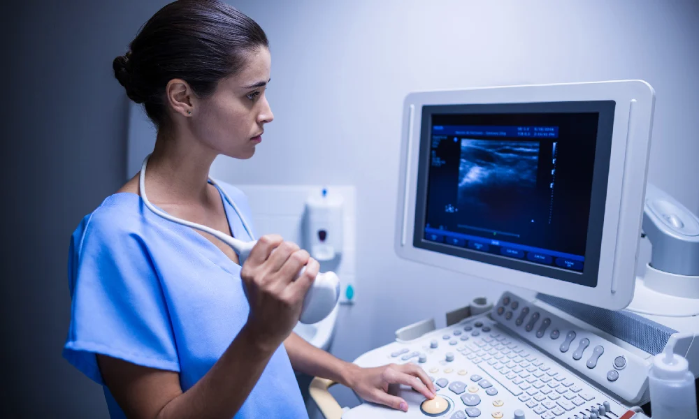
2D transesophageal echocardiography (2D TEE)
This method is the most common type of echocardiography that provides two-dimensional images of the structure of the heart. Doctors use these images to evaluate the valves, cavities, and great vessels connected to the heart.
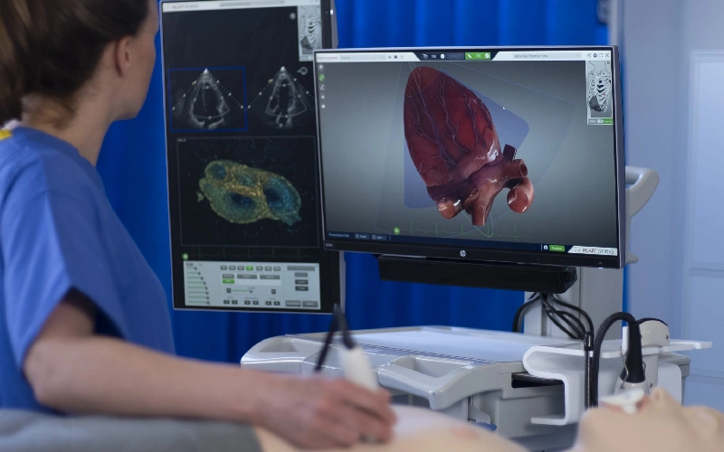
3D transesophageal echocardiography (3D TEE)
This type of echo provides three-dimensional and very detailed images of the heart, which are very useful in detecting small details such as valvular defects or heart wall defects. This type of echo is used in complex surgeries or checking the function of artificial valves.
Echo heart through Doppler (Doppler TEE)
In this method, the blood flow in the heart and main vessels is checked. This type of echo especially plays an important role in diagnosing insufficiency or stenosis of valves and hemodynamic disorders.
Stress TEE
In certain situations, echocardiography is performed under the influence of stress (pharmacological or physical) to further examine the heart's function during exercise. This method is used to evaluate coronary artery disease or evaluation before important surgeries.
Contrast TEE
With the injection of contrast material, more details of the structure of the heart and blood vessels are displayed. This method is very effective for identifying blood clots or complex heart abnormalities.
Comparison of echocardiography through the esophagus with echocardiography through the chest
|
property |
Echo through the chest |
Echo by Mary |
|
Image clarity |
Medium |
very high |
|
Equipment required |
Simple |
more advanced |
|
Need special preparation |
none |
Compulsory fasting |
| specialized applications | limited |
wider and more specialized |
Familiarity with the steps and how to perform echocardiography through the esophagus
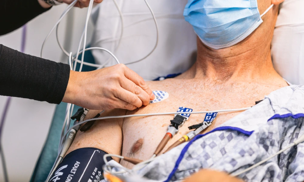
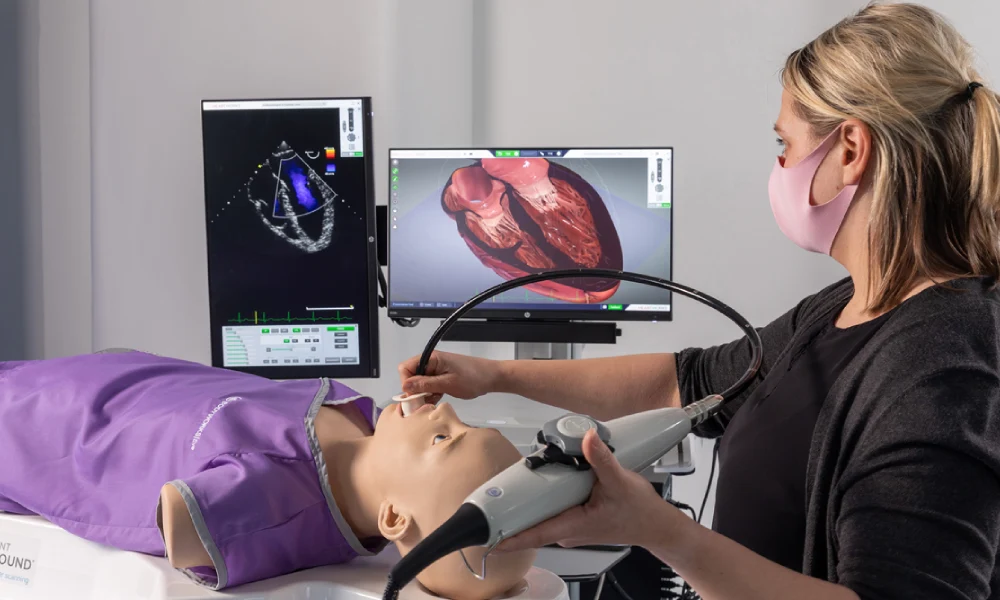
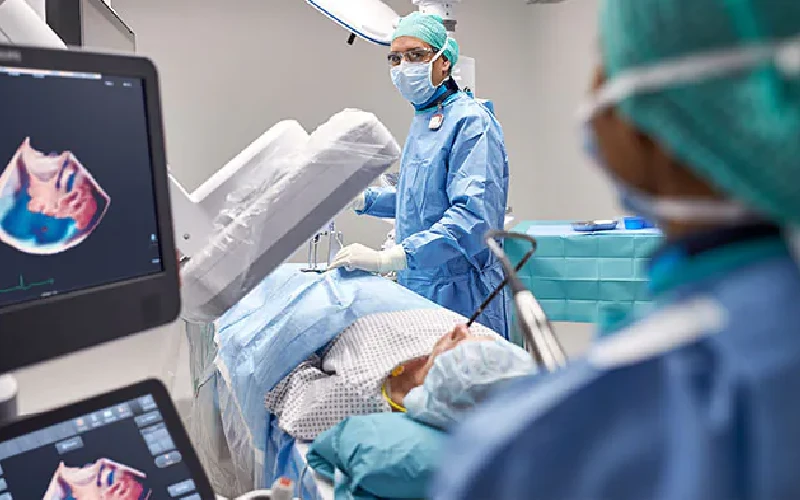
In this method, the transducer of the echocardiography device reaches near the heart through the mouth and esophagus to record clearer images of the inner parts of the heart. Doing this method includes several main steps:
- First, the patient should fast for at least 6 hours before the echo to reduce the risk of vomiting and side effects. Before starting, the doctor prepares the throat with a local anesthetic spray so that the insertion of the transducer does not cause discomfort. In most cases, a sedative is injected to keep the patient comfortable during the procedure.
- During the echo, the patient lies on his side and the transducer is slowly inserted into the esophagus through the mouth. The doctor places the transducer in the right position to capture detailed images of the heart and surrounding structures. These images are obtained by moving the transducer in different directions and changing its angle. The entire imaging process usually takes between 20 and 40 minutes.
- When finished, the converter will slowly exit. The patient may experience a mild sore throat that resolves within a few hours. It is usually recommended that the patient avoids eating and drinking until the effect of anesthesia is completely removed.
Investigation of possible complications caused by echocardiography through the esophagus
Transesophageal echocardiography (TEE) is generally a safe and low-risk procedure, but like any medical procedure, it may have risks and complications. One of the common side effects of this method is a sore throat or a feeling of discomfort in the throat after inserting the mask, which is usually temporary and resolves very quickly on the first day. In rare cases, especially if the patient has underlying problems, it may cause serious damage:
- Reactions to sedatives or local anesthesia (nausea, dizziness or allergies)
- Esophageal rupture
- internal bleeding
- Disturbance in breathing
Note that these complications occur more often in patients with complex health conditions or when referring to inexperienced doctors.
To reduce these risks, it is important for the patient to share all their medical records and drug sensitivities with the doctor before performing this procedure. Also, performing this procedure in specialized centers and under the supervision of experienced doctors can maximize its safety. In general, the benefits of this method in the accurate diagnosis of heart diseases far outweigh its risks.
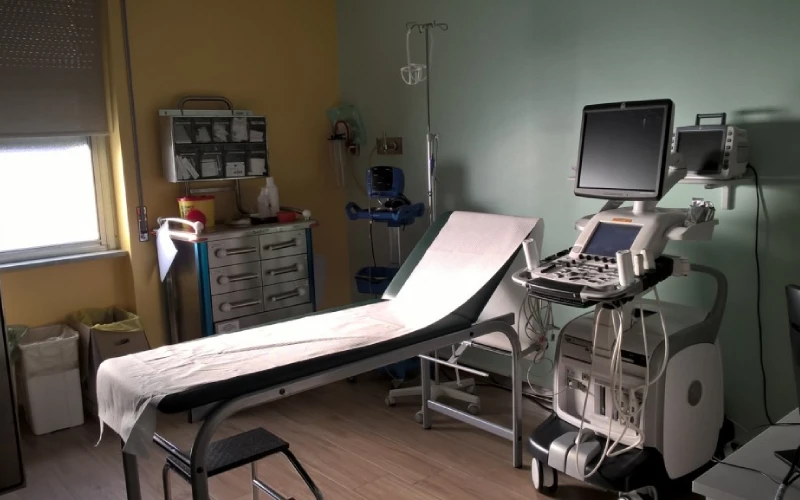
General conditions of the patient during echocardiogram test
During the test, the patient usually lies flat on the bed and carefully maintains their body position to insert the transducer into the esophagus. During the time when the doctor or echo technician inserts the transducer through the mouth into the esophagus and places it in the right position, the patient must be fully cooperative and especially calm when moving the transducer in the esophagus. The patient may have an unpleasant sensation such as a temporary sore throat or a feeling of fullness in the throat, but these discomforts are usually temporary and minor. After the test, the patient usually needs a short rest to wear off the sedative effects and return to normal. The general condition of the patient should be under the supervision of a specialist doctor so that any side effects can be identified and treated in time.
How long does a transesophageal echocardiogram take?
The duration of esophageal echocardiography is usually between 20 to 40 minutes. However, the entire process from the patient's entry into the echo room to the preparation and relaxation after the procedure may take about 1 to 2 hours.
- The initial stage takes about 10 to 15 minutes (initial preparation of the patient including checking vital signs, injecting sedatives if needed and using anesthetic spray to numb the throat)
- The second stage takes between 20 to 30 minutes depending on the complexity of the patient's condition (inserting the echo transducer into the esophagus through the mouth and imaging the heart)
After the imaging is finished, the transducer is slowly removed and the patient is monitored for a short time to wear off the sedatives.
What is the recovery period after echo through the esophagus?
The recovery period after transesophageal echocardiography (TEE) is usually short and without serious complications. This method is generally minimally invasive and the patient can return to his normal activities after a few hours.
Overall, the recovery period of this procedure is very short and most patients can return home and resume their normal activities on the same day.
People who have received a sedative should avoid driving and doing activities that require concentration for the next day. Also, on the day of the echo, it is better for the patient to be under the supervision of another person to receive help in case of an unexpected problem.
Getting to know 6 advantages of echocardiography compared to other methods
- ability to diagnose complex diseases such as blood clots in the left atrium
- ability to diagnose heart infections (endocarditis)
- Ability to detect structural abnormalities of the heart
- providing the possibility of checking the function of heart valves and valve prostheses
- essential use in cases where cardiac surgery requires detailed information
- providing reliable results in patients with excessive weight or lung problems

How much does Echo Mary cost?
The cost of echocardiography depends on various factors and may vary in different medical centers. Some of the most important factors that affect the cost of this test are:
- Type of treatment center: Usual echoes in public hospitals are usually less expensive than in private centers. Specialized heart centers may also have different costs.
- Insurance type: Many basic and supplementary insurances cover part of the cost of Echo Mary. The amount of insurance coverage depends on the type of contract and the limit of the insured's obligations.
- Equipment and technology used: Centers using more advanced and newer devices may charge more.
- Receive ancillary services: If other tests or services such as sedation are provided along with echocardiography, the final cost will increase.
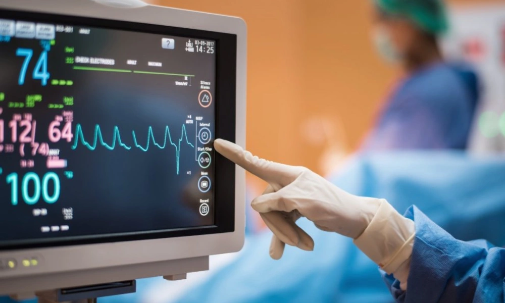
Factors that are considered in the interpretation of echocardiogram results
Interpretation of echocardiogram results can provide important information about your cardiovascular health. In an echocardiogram, images of the heart and valves are taken so that the doctor can accurately assess the heart's function, blood flow, and the condition of the vessels
.In interpreting the result of echocardiography, the first thing that is checked is the size and shape of the heart. Changes in the size of the heart chambers can be a sign of problems such as heart failure or abnormal heart enlargement. Also, the condition and function of heart valves are evaluated. Valvular diseases, such as stenosis or insufficiency of the valves, which lead to impaired blood flow, are usually detected in this test.
Another important aspect of echocardiographic interpretation is the examination of heart muscle function. If the heart muscle is weak or does not contract properly, it may be a sign of conditions such as previous heart attacks or heart failure. Also, the blood flow in large veins and arteries is also checked. Impaired blood flow or the presence of blood clots can lead to serious problems such as heart attack or stroke. Finally, the images obtained from the echo esophagus can help identify congenital heart defects, such as a hole between the two chambers of the heart.
How long does it take for transesophageal echocardiography results to be ready?
For emergencies, such as detecting blood clots or checking for a heart infection, results are usually available immediately. In non-emergency cases, a formal report including a complete analysis and treatment recommendations will usually be prepared within 24 to 48 hours.
In most cases, the initial results can be interpreted immediately after the echo is performed by a specialist doctor. During the echocardiogram, the doctor views the images live and may provide you with basic information right away.
Although the initial analysis is quick, in some cases the recorded images may require a more detailed review by a cardiologist or radiologist. This process can take a few hours to a few days, especially if the images are referred to other doctors for more detailed review.